近日浙江農(nóng)林大學(xué)在《Frontiers in Plant Science》上發(fā)表文獻《Highly efficient hairy root genetic transformation and applications in citrus》,文獻里面實驗采用了LUYOR-3415RG用于觀察GFP在毛狀根的上的表達。文獻摘要如下:
Agrobacterium-infiltrated citrus hairy root transformation
Recombinant A. rhizogenes strains were cultured in fresh yeast extract peptone medium with appropriate antibiotics at 28°C. The resuspended A. rhizogenes K599 cells at the final concentration (OD600 = 0.6) were diluted into the MES solution (10 mM MgCl2, 10 mM MES [pH 5.6], and 200 μM AS). Blade-removed citrus branches (approximately 2 months old) were collected from the greenhouse. We cut the stems into ~10 cm sections using sterilized shears by keeping the smooth surface of the cross-section.
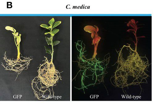
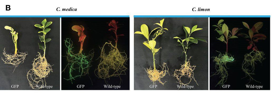
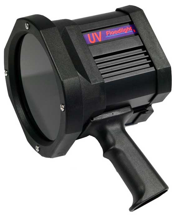
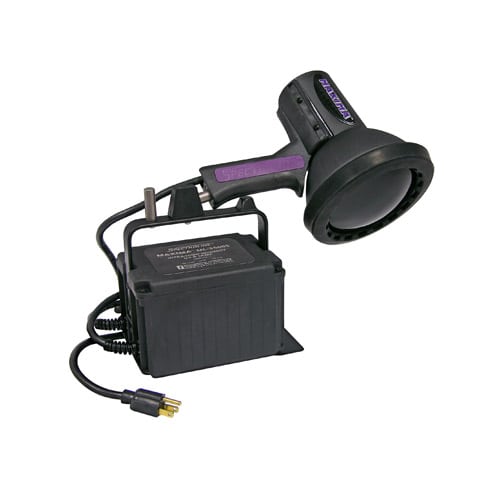
The base of the stems sections was soaked in the A. rhizogenes K599 suspension and vacuum infiltrated for approximately 25 min using a standard vacuum. The stem sections were cultured in a dome tray filled with vermiculite-mixed soil in the greenhouse at 26°C with 90% relative humidity and a 16 h/8 h (light/dark) photoperiod. Hairy root development began after approximately 2–4 weeks (C. medica) or 4–8 weeks (C. limon and citrange ‘Carrizo’) after agroinfiltrated transformation. Potential transgenic roots were detected by the fluorescence signal with a portable excitation lamp (Luyor-3415RG, Shanghai, China). The fluorescence-positive hairy roots were incubated in liquid modified Hoagland’s nutrient medium (Coolaber, Beijing, China) in sterile tubes for observation of symptoms. Transgenic roots were cultured at 26°C under a 16 h/8 h (light/dark) photoperiod. The symptoms were captured with a digital camera (Canon EOS 200D, Tokyo, Japan). Those shoots were further confirmed by PCR analysis. The hairy root transformation efficiency was calculated using the following formula: [(Number of GFP-containing roots)/(Total number of roots)] × 100. The GFP fluorescence in transgenic citrus roots was observed with a confocal microscope (LSM 780, Carl Zeiss, Jena, Germany) with 488 nm excitation and 505–530 nm emission wavelengths.
Visualization of GFP fluorescent signal
Transgenic roots were first confirmed with a hand-held excitation lamp (Luyor-3415RG). For microscopic inspection, roots were rinsed with ddH2O and photographed under a Leica-M205FA stereomicroscope (Leica Microsystems, Wetzlar, Germany). Two different autofluorescence emission wavelength bands were used for detection: green (505–550 nm) and red (>560 nm), defined by optical filters. Transverse sections of the transgenic roots were cut with a razor blade as thinly as possible. The sections were mounted on a glass microscope slide in ddH2O. The GFP signal was observed under a Leica-SP8MP confocal fluorescence microscope (Leica Microsystems). The fluorescence was observed under excitation at 488 nm and emission at 505–550 nm. Simultaneously, images (1920 × 1024 pixels) were captured with a suitable scale bar.


 添加微信咨詢!